
Diagnostic Services
Non-Invasive Cardiology
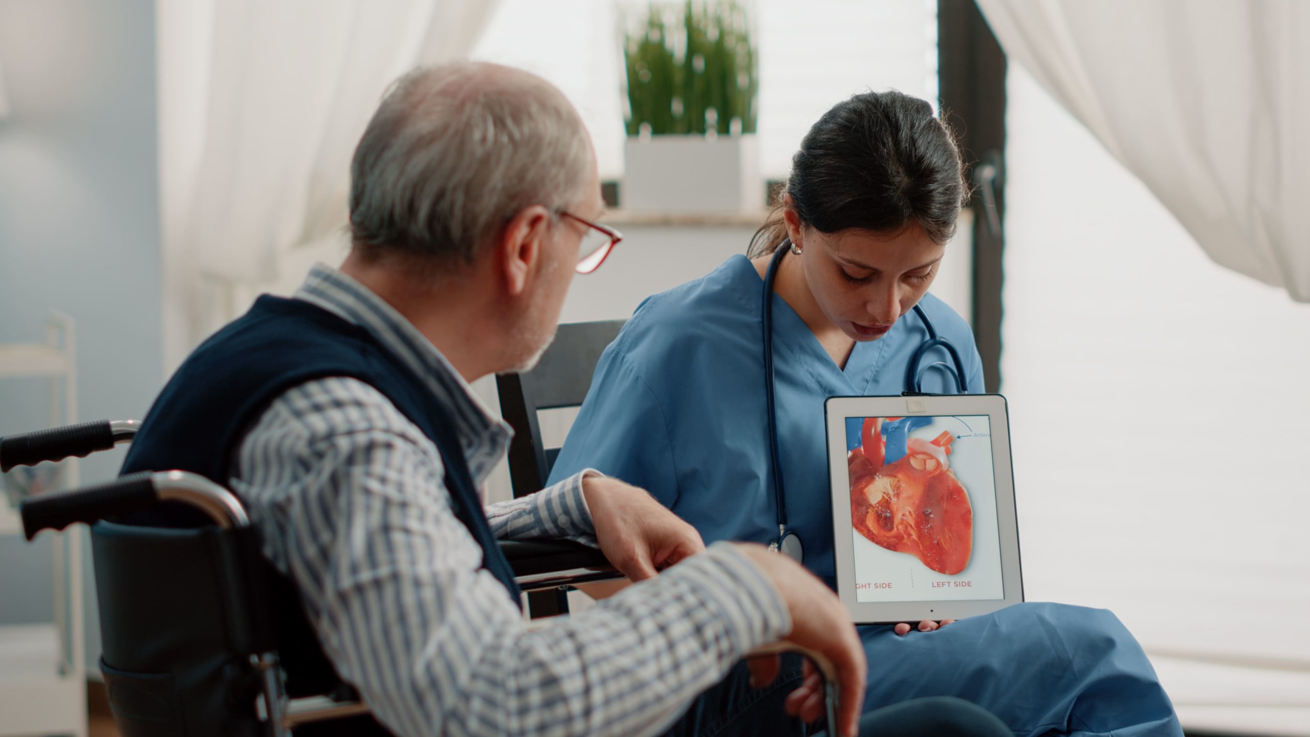
Diagnosing heart conditions effectively is the first step toward treatment and long-term health. While the heart is a complex internal organ, modern medicine allows doctors to evaluate its function and structure without the need for needles, instruments, or fluids inserted into the body. Non-invasive cardiology focuses on the detection and management of heart disorders using external tests that are safe, painless, and highly effective at identifying potential issues before they become life-threatening emergencies.
This field utilizes advanced technology to “see” the heart and monitor its activity from the outside. The primary goal is to assess the heart’s pumping ability, the condition of the valves, and the presence of any blockages in the blood supply. By using methods such as sound waves, magnetic fields, and electrical recording, cardiologists can gather critical data about the heart’s health. This approach is often the first line of defense, used to investigate symptoms like chest pain, shortness of breath, or palpitations, and to determine if more invasive procedures are actually necessary.
Two of the most common non-invasive procedures are the Echocardiogram and the Cardiac Stress Test. An Echocardiogram (or “Echo”) uses high-frequency sound waves to create moving pictures of the heart, similar to an ultrasound used during pregnancy. It allows the doctor to visualize the heart beating and pumping blood to ensure the valves are closing properly and the chambers are the correct size. A Stress Test (Treadmill Test) monitors the heart while it is working hard, usually during exercise. Since some heart problems, such as coronary artery blockages, are easier to diagnose when the heart is beating fast, this test helps reveal issues that might be missed while the patient is at rest. These diagnostic tools are vital for early detection, helping to prevent heart attacks and heart failure through timely medication and lifestyle changes.
Interventional Cardiology

Interventional Cardiology represents a specialized branch of heart care that serves as a bridge between medication-based therapy and open-heart surgery. While non-invasive tests can detect problems and surgery creates new pathways for blood flow, interventional cardiology focuses on repairing damaged vessels and structures from within the body using minimally invasive techniques. This approach utilizes catheters—thin, flexible tubes—that are threaded through the body’s blood vessels to the heart, usually entering through a small puncture in the wrist or groin. This eliminates the need for large surgical incisions or stopping the heart.
The most common procedure in this field is Percutaneous Coronary Intervention (PCI), commonly known as Angioplasty.
This is typically performed when the plaque buildup mentioned in diagnostic screenings has significantly narrowed an artery, restricting blood flow. During the procedure, a catheter with a small balloon at its tip is guided to the site of the blockage. Once in place, the balloon is inflated to compress the fatty plaque against the artery wall, widening the channel. To ensure the artery remains open long-term, a small wire mesh tube called a stent is usually placed at the site. The stent acts as a permanent scaffold, supporting the artery walls and preventing the vessel from narrowing again.
Beyond treating coronary artery disease, interventional cardiology has evolved to treat structural heart defects and valve problems that previously required open surgery. For instance, narrowed heart valves can sometimes be opened using balloon valvuloplasty, or even replaced entirely via a catheter. Because these procedures avoid the trauma of opening the chest, they typically result in significantly less pain, shorter hospital stays, and a much faster return to normal daily activities compared to traditional cardiac surgery.
Cardiac Surgery

While many heart conditions can be managed with medication or lifestyle changes, there are times when the physical structure of the heart requires surgical repair. Cardiac surgery is a specialized field focused on correcting complex issues within the heart muscle, valves, arteries, or the aorta that cannot be treated by non-invasive means alone. These procedures are performed by highly trained cardiac surgeons and are often life-saving interventions designed to restore the heart’s ability to pump blood efficiently to the rest of the body.
The most common form of cardiac surgery is Coronary Artery Bypass Grafting (CABG), often referred to simply as “bypass surgery.” This procedure is typically recommended for patients with severe Coronary Artery Disease (CAD) where multiple arteries are blocked or the blockages are too complex for stents. During this surgery, the surgeon takes a healthy blood vessel from another part of the body—usually the leg, arm, or chest—and grafts it onto the heart. This creates a new pathway, or a “detour,” around the blocked portion of the artery, allowing oxygen-rich blood to bypass the obstruction and reach the heart muscle freely.
Another critical area of cardiac surgery involves the repair or replacement of heart valves. The heart has four valves that act as one-way doors, ensuring blood flows in the correct direction. If these valves become narrowed (stenosis) and cannot open fully, or if they become leaky (regurgitation) and do not close tightly, the heart must work much harder to pump blood. Surgeons can often repair the patient’s own valve to restore its function. If repair is not possible, the damaged valve can be replaced with a prosthetic one, which may be mechanical (made of durable materials) or biological (made from animal tissue). These surgeries relieve the strain on the heart, prevent heart failure, and significantly improve the patient’s quality of life.
Preventive Cardiology
While the other branches of cardiology focus on treating existing heart conditions, Preventive Cardiology is dedicated to stopping heart disease before it starts or preventing it from worsening. It operates on the philosophy that the best way to treat a heart attack is to ensure it never happens in the first place. This field focuses on the early identification and management of risk factors that contribute to the development of Coronary Artery Disease (CAD) and other cardiovascular issues. It is particularly vital for individuals with a strong family history of heart disease or those who have already shown early warning signs.
The process begins with a comprehensive risk assessment. Since atherosclerosis (plaque buildup) is a slow, progressive disease that often begins years before symptoms appear, preventive cardiologists look for “silent” contributors. These include high blood pressure (hypertension), high cholesterol (dyslipidemia), diabetes, obesity, and smoking. By analyzing these factors alongside genetic predispositions, doctors can calculate a patient’s future risk of a cardiovascular event.
Advanced screening methods, such as calcium scoring scans or specialized blood tests, may be used to detect the earliest traces of plaque in the arteries long before a stress test would show any abnormalities.
Once risks are identified, the focus shifts to aggressive management through a combination of lifestyle modification and medical therapy. This is not a “one-size-fits-all” approach but a personalized strategy. It may involve nutritional counseling to lower cholesterol, tailored exercise programs to strengthen the heart, and smoking cessation support. When lifestyle changes alone are insufficient, medications such as statins (to lower cholesterol) or anti-hypertensives are prescribed to protect the vessel walls. By controlling these variables, Preventive Cardiology aims to halt the progression of plaque, keep the arteries flexible and clear, and ensure a longer, healthier life free from the complications of advanced heart disease.
Electrophysiology
Just as a house needs electrical wiring to power its lights, the heart relies on a complex internal electrical system to regulate its rhythm. This system generates the signals that tell the heart muscle when to contract and pump blood. Clinical Electrophysiology (EP) is the specialized branch of cardiology dedicated to diagnosing and treating disorders of this electrical system, known as arrhythmias. When the electrical signals malfunction, the heart may beat too fast (tachycardia), too slow (bradycardia), or irregularly (such as in Atrial Fibrillation). These irregularities can cause symptoms ranging from palpitations and dizziness to fainting or even sudden cardiac arrest.
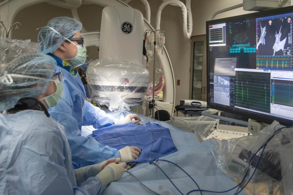
To diagnose these complex rhythm disorders, doctors often perform an Electrophysiology (EP) Study. This is a minimally invasive procedure where specialized wire electrodes are guided into the heart through blood vessels, similar to the technique used in angioplasty. These electrodes allow the electrophysiologist to “map” the heart’s electrical activity with high precision. By recording the electrical signals from inside the heart, they can locate the exact source of the “short circuit” or abnormal pathway that is disturbing the natural rhythm.
Once the specific issue is identified, it can often be treated or even cured during the same procedure. A common treatment is Radiofrequency Ablation, where heat energy is used to carefully neutralize the tiny area of tissue causing the abnormal signal, restoring normal rhythm without the need for open surgery. For patients whose hearts beat too slowly or are at risk of stopping, implantable devices are used. A Pacemaker is a small device implanted under the skin that sends electrical pulses to prompt the heart to beat at a normal rate, while an Implantable Cardioverter Defibrillator (ICD) monitors the heart 24/7 and can deliver a life-saving shock if it detects a dangerous, rapid rhythm.
Non-Invasive Cardiology
Diagnosing heart conditions effectively is the first step toward treatment and long-term health. While the heart is a complex internal organ, modern medicine allows doctors to evaluate its function and structure without the need for needles, instruments, or fluids inserted into the body. Non-invasive cardiology focuses on the detection and management of heart disorders using external tests that are safe, painless, and highly effective at identifying potential issues before they become life-threatening emergencies.
This field utilizes advanced technology to “see” the heart and monitor its activity from the outside. The primary goal is to assess the heart’s pumping ability, the condition of the valves, and the presence of any blockages in the blood supply. By using methods such as sound waves, magnetic fields, and electrical recording, cardiologists can gather critical data about the heart’s health. This approach is often the first line of defense, used to investigate symptoms like chest pain, shortness of breath, or palpitations, and to determine if more invasive procedures are actually necessary.
Two of the most common non-invasive procedures are the Echocardiogram and the Cardiac Stress Test. An Echocardiogram (or “Echo”) uses high-frequency sound waves to create moving pictures of the heart, similar to an ultrasound used during pregnancy. It allows the doctor to visualize the heart beating and pumping blood to ensure the valves are closing properly and the chambers are the correct size. A Stress Test (Treadmill Test) monitors the heart while it is working hard, usually during exercise. Since some heart problems, such as coronary artery blockages, are easier to diagnose when the heart is beating fast, this test helps reveal issues that might be missed while the patient is at rest. These diagnostic tools are vital for early detection, helping to prevent heart attacks and heart failure through timely medication and lifestyle changes.
Interventional Cardiology
Interventional Cardiology represents a specialized branch of heart care that serves as a bridge between medication-based therapy and open-heart surgery. While non-invasive tests can detect problems and surgery creates new pathways for blood flow, interventional cardiology focuses on repairing damaged vessels and structures from within the body using minimally invasive techniques. This approach utilizes catheters—thin, flexible tubes—that are threaded through the body’s blood vessels to the heart, usually entering through a small puncture in the wrist or groin. This eliminates the need for large surgical incisions or stopping the heart.
The most common procedure in this field is Percutaneous Coronary Intervention (PCI), commonly known as Angioplasty.
This is typically performed when the plaque buildup mentioned in diagnostic screenings has significantly narrowed an artery, restricting blood flow. During the procedure, a catheter with a small balloon at its tip is guided to the site of the blockage. Once in place, the balloon is inflated to compress the fatty plaque against the artery wall, widening the channel. To ensure the artery remains open long-term, a small wire mesh tube called a stent is usually placed at the site. The stent acts as a permanent scaffold, supporting the artery walls and preventing the vessel from narrowing again.
Beyond treating coronary artery disease, interventional cardiology has evolved to treat structural heart defects and valve problems that previously required open surgery. For instance, narrowed heart valves can sometimes be opened using balloon valvuloplasty, or even replaced entirely via a catheter. Because these procedures avoid the trauma of opening the chest, they typically result in significantly less pain, shorter hospital stays, and a much faster return to normal daily activities compared to traditional cardiac surgery.
Cardiac Surgery
While many heart conditions can be managed with medication or lifestyle changes, there are times when the physical structure of the heart requires surgical repair. Cardiac surgery is a specialized field focused on correcting complex issues within the heart muscle, valves, arteries, or the aorta that cannot be treated by non-invasive means alone. These procedures are performed by highly trained cardiac surgeons and are often life-saving interventions designed to restore the heart’s ability to pump blood efficiently to the rest of the body.
The most common form of cardiac surgery is Coronary Artery Bypass Grafting (CABG), often referred to simply as “bypass surgery.” This procedure is typically recommended for patients with severe Coronary Artery Disease (CAD) where multiple arteries are blocked or the blockages are too complex for stents. During this surgery, the surgeon takes a healthy blood vessel from another part of the body—usually the leg, arm, or chest—and grafts it onto the heart. This creates a new pathway, or a “detour,” around the blocked portion of the artery, allowing oxygen-rich blood to bypass the obstruction and reach the heart muscle freely.
Another critical area of cardiac surgery involves the repair or replacement of heart valves. The heart has four valves that act as one-way doors, ensuring blood flows in the correct direction. If these valves become narrowed (stenosis) and cannot open fully, or if they become leaky (regurgitation) and do not close tightly, the heart must work much harder to pump blood. Surgeons can often repair the patient’s own valve to restore its function. If repair is not possible, the damaged valve can be replaced with a prosthetic one, which may be mechanical (made of durable materials) or biological (made from animal tissue). These surgeries relieve the strain on the heart, prevent heart failure, and significantly improve the patient’s quality of life.
Preventive Cardiology
While the other branches of cardiology focus on treating existing heart conditions, Preventive Cardiology is dedicated to stopping heart disease before it starts or preventing it from worsening. It operates on the philosophy that the best way to treat a heart attack is to ensure it never happens in the first place. This field focuses on the early identification and management of risk factors that contribute to the development of Coronary Artery Disease (CAD) and other cardiovascular issues. It is particularly vital for individuals with a strong family history of heart disease or those who have already shown early warning signs.
The process begins with a comprehensive risk assessment. Since atherosclerosis (plaque buildup) is a slow, progressive disease that often begins years before symptoms appear, preventive cardiologists look for “silent” contributors. These include high blood pressure (hypertension), high cholesterol (dyslipidemia), diabetes, obesity, and smoking. By analyzing these factors alongside genetic predispositions, doctors can calculate a patient’s future risk of a cardiovascular event.
Advanced screening methods, such as calcium scoring scans or specialized blood tests, may be used to detect the earliest traces of plaque in the arteries long before a stress test would show any abnormalities.
Once risks are identified, the focus shifts to aggressive management through a combination of lifestyle modification and medical therapy. This is not a “one-size-fits-all” approach but a personalized strategy. It may involve nutritional counseling to lower cholesterol, tailored exercise programs to strengthen the heart, and smoking cessation support. When lifestyle changes alone are insufficient, medications such as statins (to lower cholesterol) or anti-hypertensives are prescribed to protect the vessel walls. By controlling these variables, Preventive Cardiology aims to halt the progression of plaque, keep the arteries flexible and clear, and ensure a longer, healthier life free from the complications of advanced heart disease.
Electrophysiology
Just as a house needs electrical wiring to power its lights, the heart relies on a complex internal electrical system to regulate its rhythm. This system generates the signals that tell the heart muscle when to contract and pump blood. Clinical Electrophysiology (EP) is the specialized branch of cardiology dedicated to diagnosing and treating disorders of this electrical system, known as arrhythmias. When the electrical signals malfunction, the heart may beat too fast (tachycardia), too slow (bradycardia), or irregularly (such as in Atrial Fibrillation). These irregularities can cause symptoms ranging from palpitations and dizziness to fainting or even sudden cardiac arrest.
To diagnose these complex rhythm disorders, doctors often perform an Electrophysiology (EP) Study. This is a minimally invasive procedure where specialized wire electrodes are guided into the heart through blood vessels, similar to the technique used in angioplasty. These electrodes allow the electrophysiologist to “map” the heart’s electrical activity with high precision. By recording the electrical signals from inside the heart, they can locate the exact source of the “short circuit” or abnormal pathway that is disturbing the natural rhythm.
Once the specific issue is identified, it can often be treated or even cured during the same procedure. A common treatment is Radiofrequency Ablation, where heat energy is used to carefully neutralize the tiny area of tissue causing the abnormal signal, restoring normal rhythm without the need for open surgery. For patients whose hearts beat too slowly or are at risk of stopping, implantable devices are used. A Pacemaker is a small device implanted under the skin that sends electrical pulses to prompt the heart to beat at a normal rate, while an Implantable Cardioverter Defibrillator (ICD) monitors the heart 24/7 and can deliver a life-saving shock if it detects a dangerous, rapid rhythm.
Emergency Care

Cardiac emergencies are unpredictable and require immediate medical attention to save lives and minimize permanent heart damage. Unlike scheduled check-ups or elective surgeries, emergency cardiac care operates on the principle that “time is muscle.” When blood flow to the heart is blocked, heart muscle begins to die within minutes. Therefore, the primary goal of emergency care is rapid stabilization, quick diagnosis, and the immediate restoration of blood flow to prevent long-term complications or fatality.
The most critical condition managed in this setting is the Acute Myocardial Infarction, or heart attack. Medical professionals often refer to the “Golden Hour”—the first hour after the onset of severe symptoms—as the most crucial window for treatment. During this time, immediate interventions can often reverse the blockage before irreversible damage occurs. Patients presenting with symptoms such as crushing chest pain, profuse sweating, difficulty breathing, or sudden loss of consciousness are prioritized instantly. Specialized teams are on standby 24/7 to perform urgent diagnostic tests like an ECG to identify the exact nature of the emergency.
Modern emergency cardiac care often involves “Primary Angioplasty,” which is considered the gold standard for treating major heart attacks. Instead of waiting for clot-busting medicines to work, the patient is rushed directly to the catheterization laboratory. Here, interventional cardiologists mechanically open the blocked artery using a balloon and stent. This seamless coordination between the emergency room and the cardiac team ensures that the time from the patient entering the hospital door to the opening of the artery is kept as short as possible, significantly increasing the chances of survival and a full recovery.
Intensive Care

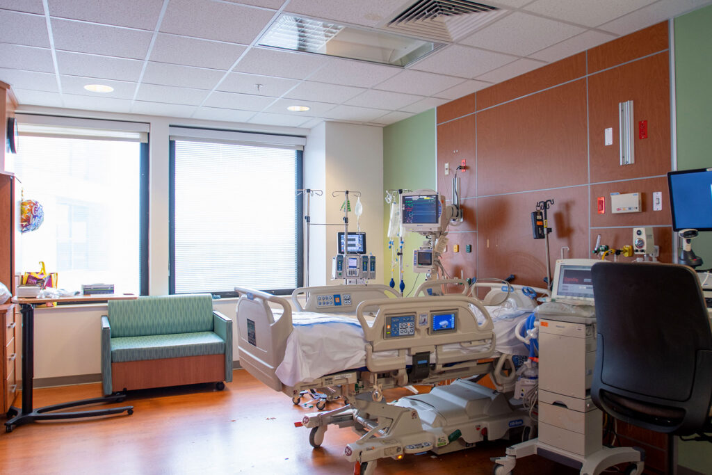
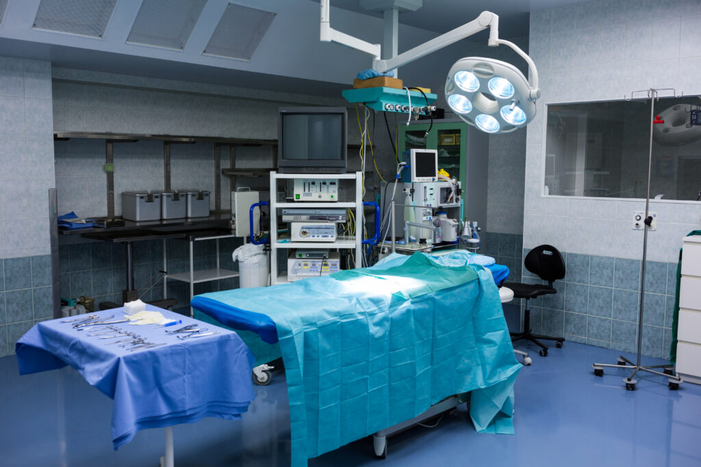
The Intensive Care Unit (ICU) represents the pinnacle of medical monitoring and support within a hospital. It is a specialized department dedicated to the management of patients with life-threatening illnesses, severe injuries, or those recovering from complex surgeries who require constant, close attention. Unlike a standard hospital ward, the ICU provides a highly controlled environment where every aspect of a patient’s condition—from heart rate and blood pressure to oxygen levels and organ function—is observed continuously, minute by minute.
This level of care is made possible by a combination of advanced technology and life-support systems. Patients in the ICU are connected to sophisticated bedside monitors that feed real-time data to a central nursing station, ensuring that the medical team is alerted instantly to even the slightest change in vital signs. For patients who cannot breathe effectively on their own, mechanical ventilators are used to assist or take over respiration. Other specialized equipment may be used to support the heart, kidneys, or circulatory system, stabilizing the body’s vital functions to buy time for the underlying condition to be treated and healed.
However, the true strength of the Intensive Care Unit lies in its specialized team. The unit is led by Intensivists—doctors with advanced training in critical care medicine—who are available around the clock to make immediate, complex decisions. They work alongside critical care nurses who provide a very high level of bedside care, often on a one-to-one basis with the patient. This multidisciplinary team, which often includes respiratory therapists, nutritionists, and physiotherapists, works in unison to guide patients through the most fragile phase of their recovery, aiming to restore stability and transition them back to normal ward care.
Pathology

Pathology is often described as the “hidden engine” of a hospital. While patients spend their time interacting with physicians and surgeons, the course of their treatment is frequently determined by the scientific analysis that happens behind the scenes in the pathology laboratory. This department is dedicated to the study of disease through the examination of bodily fluids, tissues, and organs. By analyzing samples—most commonly blood, urine, or biopsy tissue—pathologists provide the objective data that doctors rely on to make accurate diagnoses, monitor the progress of chronic conditions, and verify that treatments are working effectively.
The majority of routine testing falls under Clinical Pathology and Biochemistry. These areas focus on analyzing the chemical components and cellular makeup of blood and other fluids. Whether it is checking cholesterol levels to assess heart risk, monitoring blood sugar for diabetes management, or evaluating liver and kidney function, these tests provide a comprehensive “snapshot” of the body’s internal health. Advanced automated analyzers are used to process these samples with high precision and speed, ensuring that critical results are available to clinicians as quickly as possible, often within hours.
For more complex diagnostic challenges, Histopathology and Cytology are essential. In these procedures, pathologists examine tissue samples (biopsies) or cell clusters under a microscope to look for structural abnormalities at a cellular level. This is the gold standard for diagnosing cancers, tumors, and specific inflammatory conditions. The pathologist acts as a “doctor’s doctor,” interpreting these intricate findings to differentiate between benign (harmless) and malignant (cancerous) growths. This definitive diagnosis allows the treating physician to tailor a therapy plan that addresses the specific nature of the disease.
Pulmonology
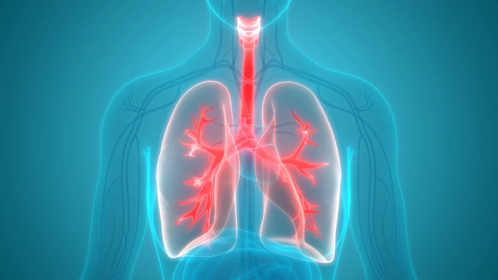
Breathing is the most fundamental act of life, yet it is often taken for granted until it becomes difficult. Pulmonology is the specialized branch of medicine dedicated to the health of the respiratory system, specifically the lungs, windpipe, and bronchial tubes. This field focuses on ensuring that the body can effectively take in oxygen and expel carbon dioxide, a process vital for the function of every cell. Pulmonologists are experts in diagnosing and treating conditions that affect breathing, ranging from temporary infections to chronic, long-term respiratory disorders.
A significant portion of pulmonology involves managing chronic conditions such as Asthma, Chronic Obstructive Pulmonary Disease (COPD), and Interstitial Lung Disease. Diagnosis often begins with a Pulmonary Function Test (PFT) or Spirometry. This non-invasive test measures the volume and speed of air a patient can inhale and exhale, providing a clear picture of lung capacity and health. For more detailed investigation, doctors may use Bronchoscopy, a procedure where a thin, flexible tube with a camera is passed gently down the throat to directly visualize the airways. This allows physicians to check for blockages, infections, or tumors without the need for surgery.
Beyond treating lung diseases, Pulmonology also plays a critical role in Sleep Medicine, particularly in the diagnosis and management of Obstructive Sleep Apnea—a condition where breathing repeatedly stops and starts during sleep. Treatment plans are highly personalized and may involve medication, inhalers, pulmonary rehabilitation exercises, or the use of devices like CPAP machines to keep airways open at night. The ultimate goal is to help patients breathe easier, improve their oxygen levels, and restore their stamina and quality of life.
Nephrology Service
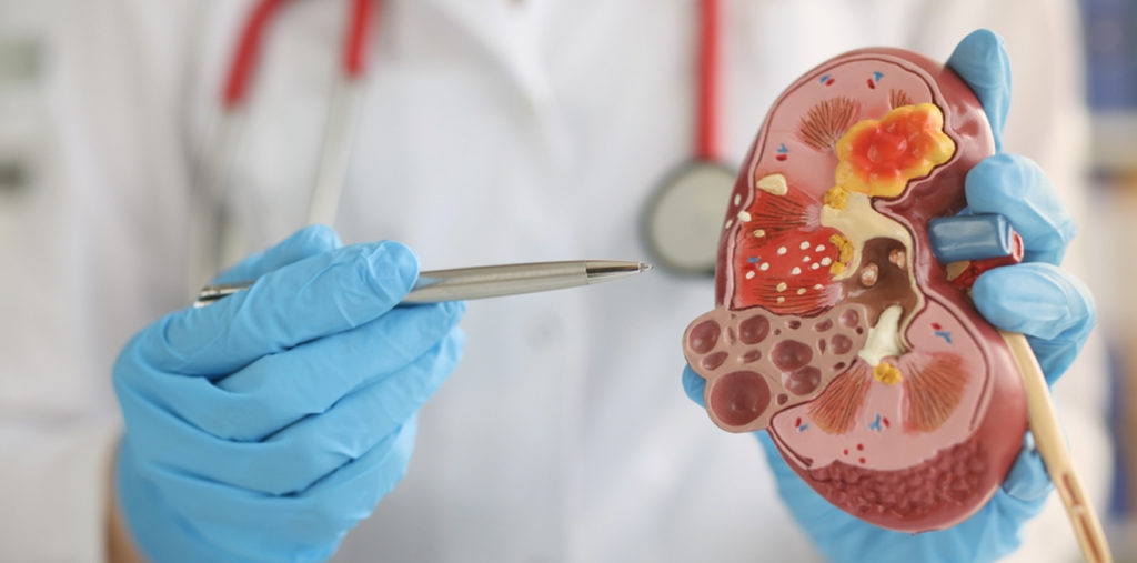
The kidneys are often referred to as the body’s master chemists. These two bean-shaped organs, located on either side of the spine, perform the vital task of filtering blood to remove toxins, excess fluids, and waste products, which are then excreted as urine. Beyond filtration, they also regulate blood pressure, balance electrolytes, and produce hormones that keep bones strong and blood healthy. Nephrology is the specialized branch of internal medicine dedicated to the study, diagnosis, and treatment of kidney diseases, ensuring these complex filtration systems continue to function effectively.
A significant part of nephrology focuses on the management of Chronic Kidney Disease (CKD), a condition where kidney function gradually declines over time. This is often caused by long-standing diabetes or high blood pressure (hypertension). Nephrologists utilize precise blood and urine tests to monitor the “Glomerular Filtration Rate” (GFR)—a measure of how well the kidneys are cleaning the blood. Early detection is crucial; by identifying kidney stress early, doctors can prescribe medications and dietary changes to slow the progression of disease, prevent the formation of kidney stones, and correct electrolyte imbalances before they become critical.
When the kidneys are no longer able to function on their own (End-Stage Renal Disease), nephrology services provide life-sustaining treatments through Dialysis. Dialysis acts as an artificial kidney, mechanically filtering the blood to keep the patient alive. This can be done via Haemodialysis, where blood is cycled through an external machine to be cleaned and returned to the body, or Peritoneal Dialysis, which uses the lining of the abdomen to filter the blood inside the body. For eligible patients, nephrologists also manage the preparation and post-operative care for Kidney Transplantation, offering the best chance for a return to a normal lifestyle.
Diabetalogy Service
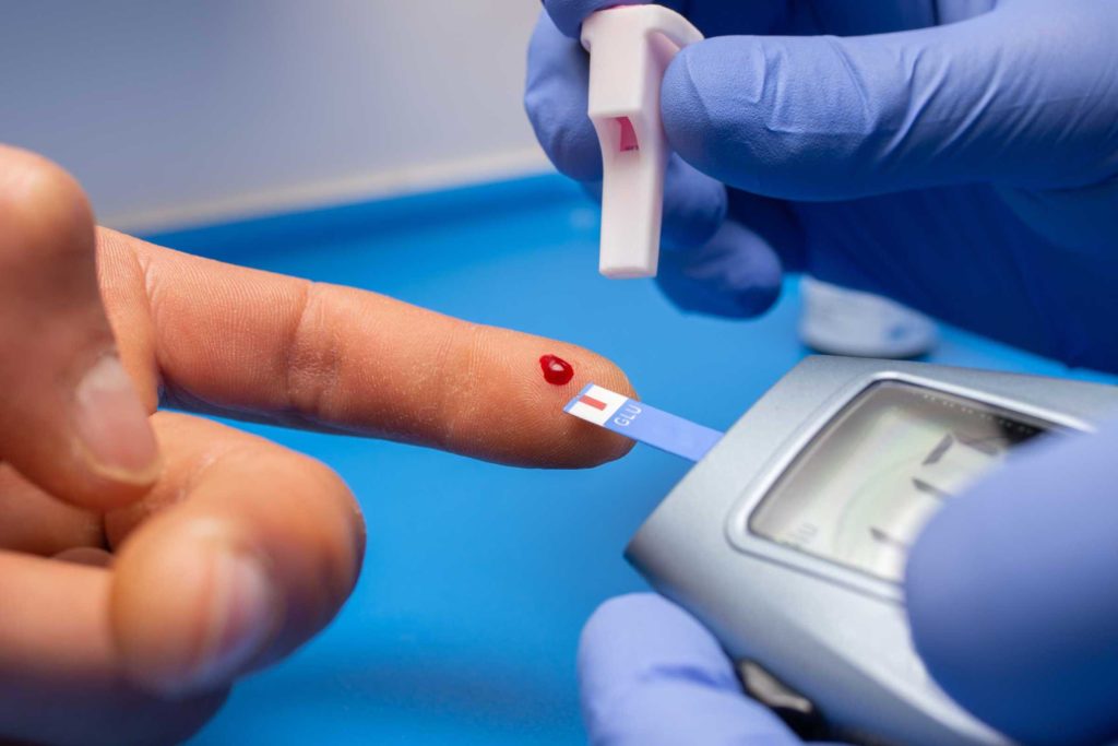
Diabetes Mellitus is often referred to as a “silent” condition because it can develop gradually over years without causing obvious symptoms. It is a chronic metabolic disorder that affects how the body utilizes glucose (blood sugar), which is the primary source of energy for the cells. The key to this process is insulin, a hormone produced by the pancreas that acts as a “key” to unlock cells and let glucose enter. Diabetology is the specialized field dedicated to diagnosing, treating, and managing this complex condition to ensure that blood sugar levels remain within a safe range.
There are two primary forms of the disease managed by this service. Type 1 Diabetes is an autoimmune reaction where the body stops producing insulin entirely, often requiring daily insulin replacement. Type 2 Diabetes, which is far more common, occurs when the body becomes resistant to the insulin it produces or doesn’t make enough. Left uncontrolled, high blood sugar acts like a slow-acting poison, damaging blood vessels and nerves throughout the body. Diabetologists use comprehensive screening tools, such as the HbA1c test (which gives a 3-month average of blood sugar control), to diagnose the condition accurately and monitor its progression.
Modern diabetes care is holistic and goes far beyond just prescribing medication. While oral drugs or insulin injections are often necessary to regulate glucose levels, the cornerstone of treatment involves lifestyle management. Diabetologists work closely with nutritionists and educators to create personalized diet plans and exercise regimes that help improve insulin sensitivity. The ultimate goal of the service is not just to lower numbers on a meter, but to prevent the serious long-term complications of diabetes, such as kidney failure, vision loss (retinopathy), and nerve damage (neuropathy), ensuring patients can lead active, healthy lives.
Non-Invasive Cardiology
Diagnosing heart conditions effectively is the first step toward treatment and long-term health. While the heart is a complex internal organ, modern medicine allows doctors to evaluate its function and structure without the need for needles, instruments, or fluids inserted into the body. Non-invasive cardiology focuses on the detection and management of heart disorders using external tests that are safe, painless, and highly effective at identifying potential issues before they become life-threatening emergencies.
This field utilizes advanced technology to “see” the heart and monitor its activity from the outside. The primary goal is to assess the heart’s pumping ability, the condition of the valves, and the presence of any blockages in the blood supply. By using methods such as sound waves, magnetic fields, and electrical recording, cardiologists can gather critical data about the heart’s health. This approach is often the first line of defense, used to investigate symptoms like chest pain, shortness of breath, or palpitations, and to determine if more invasive procedures are actually necessary.
Two of the most common non-invasive procedures are the Echocardiogram and the Cardiac Stress Test. An Echocardiogram (or “Echo”) uses high-frequency sound waves to create moving pictures of the heart, similar to an ultrasound used during pregnancy. It allows the doctor to visualize the heart beating and pumping blood to ensure the valves are closing properly and the chambers are the correct size. A Stress Test (Treadmill Test) monitors the heart while it is working hard, usually during exercise. Since some heart problems, such as coronary artery blockages, are easier to diagnose when the heart is beating fast, this test helps reveal issues that might be missed while the patient is at rest. These diagnostic tools are vital for early detection, helping to prevent heart attacks and heart failure through timely medication and lifestyle changes.
Cardiac Surgery
While many heart conditions can be managed with medication or lifestyle changes, there are times when the physical structure of the heart requires surgical repair. Cardiac surgery is a specialized field focused on correcting complex issues within the heart muscle, valves, arteries, or the aorta that cannot be treated by non-invasive means alone. These procedures are performed by highly trained cardiac surgeons and are often life-saving interventions designed to restore the heart’s ability to pump blood efficiently to the rest of the body.
The most common form of cardiac surgery is Coronary Artery Bypass Grafting (CABG), often referred to simply as “bypass surgery.” This procedure is typically recommended for patients with severe Coronary Artery Disease (CAD) where multiple arteries are blocked or the blockages are too complex for stents. During this surgery, the surgeon takes a healthy blood vessel from another part of the body—usually the leg, arm, or chest—and grafts it onto the heart. This creates a new pathway, or a “detour,” around the blocked portion of the artery, allowing oxygen-rich blood to bypass the obstruction and reach the heart muscle freely.
Another critical area of cardiac surgery involves the repair or replacement of heart valves. The heart has four valves that act as one-way doors, ensuring blood flows in the correct direction. If these valves become narrowed (stenosis) and cannot open fully, or if they become leaky (regurgitation) and do not close tightly, the heart must work much harder to pump blood. Surgeons can often repair the patient’s own valve to restore its function. If repair is not possible, the damaged valve can be replaced with a prosthetic one, which may be mechanical (made of durable materials) or biological (made from animal tissue). These surgeries relieve the strain on the heart, prevent heart failure, and significantly improve the patient’s quality of life.
Interventional Cardiology
Interventional Cardiology represents a specialized branch of heart care that serves as a bridge between medication-based therapy and open-heart surgery. While non-invasive tests can detect problems and surgery creates new pathways for blood flow, interventional cardiology focuses on repairing damaged vessels and structures from within the body using minimally invasive techniques. This approach utilizes catheters—thin, flexible tubes—that are threaded through the body’s blood vessels to the heart, usually entering through a small puncture in the wrist or groin. This eliminates the need for large surgical incisions or stopping the heart.
The most common procedure in this field is Percutaneous Coronary Intervention (PCI), commonly known as Angioplasty.
This is typically performed when the plaque buildup mentioned in diagnostic screenings has significantly narrowed an artery, restricting blood flow. During the procedure, a catheter with a small balloon at its tip is guided to the site of the blockage. Once in place, the balloon is inflated to compress the fatty plaque against the artery wall, widening the channel. To ensure the artery remains open long-term, a small wire mesh tube called a stent is usually placed at the site. The stent acts as a permanent scaffold, supporting the artery walls and preventing the vessel from narrowing again.
Beyond treating coronary artery disease, interventional cardiology has evolved to treat structural heart defects and valve problems that previously required open surgery. For instance, narrowed heart valves can sometimes be opened using balloon valvuloplasty, or even replaced entirely via a catheter. Because these procedures avoid the trauma of opening the chest, they typically result in significantly less pain, shorter hospital stays, and a much faster return to normal daily activities compared to traditional cardiac surgery.
Preventive Cardiology
While the other branches of cardiology focus on treating existing heart conditions, Preventive Cardiology is dedicated to stopping heart disease before it starts or preventing it from worsening. It operates on the philosophy that the best way to treat a heart attack is to ensure it never happens in the first place. This field focuses on the early identification and management of risk factors that contribute to the development of Coronary Artery Disease (CAD) and other cardiovascular issues. It is particularly vital for individuals with a strong family history of heart disease or those who have already shown early warning signs.
The process begins with a comprehensive risk assessment. Since atherosclerosis (plaque buildup) is a slow, progressive disease that often begins years before symptoms appear, preventive cardiologists look for “silent” contributors. These include high blood pressure (hypertension), high cholesterol (dyslipidemia), diabetes, obesity, and smoking. By analyzing these factors alongside genetic predispositions, doctors can calculate a patient’s future risk of a cardiovascular event.
Advanced screening methods, such as calcium scoring scans or specialized blood tests, may be used to detect the earliest traces of plaque in the arteries long before a stress test would show any abnormalities.
Once risks are identified, the focus shifts to aggressive management through a combination of lifestyle modification and medical therapy. This is not a “one-size-fits-all” approach but a personalized strategy. It may involve nutritional counseling to lower cholesterol, tailored exercise programs to strengthen the heart, and smoking cessation support. When lifestyle changes alone are insufficient, medications such as statins (to lower cholesterol) or anti-hypertensives are prescribed to protect the vessel walls. By controlling these variables, Preventive Cardiology aims to halt the progression of plaque, keep the arteries flexible and clear, and ensure a longer, healthier life free from the complications of advanced heart disease.
Emergency Care
Cardiac emergencies are unpredictable and require immediate medical attention to save lives and minimize permanent heart damage. Unlike scheduled check-ups or elective surgeries, emergency cardiac care operates on the principle that “time is muscle.” When blood flow to the heart is blocked, heart muscle begins to die within minutes. Therefore, the primary goal of emergency care is rapid stabilization, quick diagnosis, and the immediate restoration of blood flow to prevent long-term complications or fatality.
The most critical condition managed in this setting is the Acute Myocardial Infarction, or heart attack. Medical professionals often refer to the “Golden Hour”—the first hour after the onset of severe symptoms—as the most crucial window for treatment. During this time, immediate interventions can often reverse the blockage before irreversible damage occurs. Patients presenting with symptoms such as crushing chest pain, profuse sweating, difficulty breathing, or sudden loss of consciousness are prioritized instantly. Specialized teams are on standby 24/7 to perform urgent diagnostic tests like an ECG to identify the exact nature of the emergency.
Modern emergency cardiac care often involves “Primary Angioplasty,” which is considered the gold standard for treating major heart attacks. Instead of waiting for clot-busting medicines to work, the patient is rushed directly to the catheterization laboratory. Here, interventional cardiologists mechanically open the blocked artery using a balloon and stent. This seamless coordination between the emergency room and the cardiac team ensures that the time from the patient entering the hospital door to the opening of the artery is kept as short as possible, significantly increasing the chances of survival and a full recovery.
Electrophysiology
Just as a house needs electrical wiring to power its lights, the heart relies on a complex internal electrical system to regulate its rhythm. This system generates the signals that tell the heart muscle when to contract and pump blood. Clinical Electrophysiology (EP) is the specialized branch of cardiology dedicated to diagnosing and treating disorders of this electrical system, known as arrhythmias. When the electrical signals malfunction, the heart may beat too fast (tachycardia), too slow (bradycardia), or irregularly (such as in Atrial Fibrillation). These irregularities can cause symptoms ranging from palpitations and dizziness to fainting or even sudden cardiac arrest.
To diagnose these complex rhythm disorders, doctors often perform an Electrophysiology (EP) Study. This is a minimally invasive procedure where specialized wire electrodes are guided into the heart through blood vessels, similar to the technique used in angioplasty. These electrodes allow the electrophysiologist to “map” the heart’s electrical activity with high precision. By recording the electrical signals from inside the heart, they can locate the exact source of the “short circuit” or abnormal pathway that is disturbing the natural rhythm.
Once the specific issue is identified, it can often be treated or even cured during the same procedure. A common treatment is Radiofrequency Ablation, where heat energy is used to carefully neutralize the tiny area of tissue causing the abnormal signal, restoring normal rhythm without the need for open surgery. For patients whose hearts beat too slowly or are at risk of stopping, implantable devices are used. A Pacemaker is a small device implanted under the skin that sends electrical pulses to prompt the heart to beat at a normal rate, while an Implantable Cardioverter Defibrillator (ICD) monitors the heart 24/7 and can deliver a life-saving shock if it detects a dangerous, rapid rhythm.
Pathology
Pathology is often described as the “hidden engine” of a hospital. While patients spend their time interacting with physicians and surgeons, the course of their treatment is frequently determined by the scientific analysis that happens behind the scenes in the pathology laboratory. This department is dedicated to the study of disease through the examination of bodily fluids, tissues, and organs. By analyzing samples—most commonly blood, urine, or biopsy tissue—pathologists provide the objective data that doctors rely on to make accurate diagnoses, monitor the progress of chronic conditions, and verify that treatments are working effectively.
The majority of routine testing falls under Clinical Pathology and Biochemistry. These areas focus on analyzing the chemical components and cellular makeup of blood and other fluids. Whether it is checking cholesterol levels to assess heart risk, monitoring blood sugar for diabetes management, or evaluating liver and kidney function, these tests provide a comprehensive “snapshot” of the body’s internal health. Advanced automated analyzers are used to process these samples with high precision and speed, ensuring that critical results are available to clinicians as quickly as possible, often within hours.
For more complex diagnostic challenges, Histopathology and Cytology are essential. In these procedures, pathologists examine tissue samples (biopsies) or cell clusters under a microscope to look for structural abnormalities at a cellular level. This is the gold standard for diagnosing cancers, tumors, and specific inflammatory conditions. The pathologist acts as a “doctor’s doctor,” interpreting these intricate findings to differentiate between benign (harmless) and malignant (cancerous) growths. This definitive diagnosis allows the treating physician to tailor a therapy plan that addresses the specific nature of the disease.
Intensive Care
The Intensive Care Unit (ICU) represents the pinnacle of medical monitoring and support within a hospital. It is a specialized department dedicated to the management of patients with life-threatening illnesses, severe injuries, or those recovering from complex surgeries who require constant, close attention. Unlike a standard hospital ward, the ICU provides a highly controlled environment where every aspect of a patient’s condition—from heart rate and blood pressure to oxygen levels and organ function—is observed continuously, minute by minute.
This level of care is made possible by a combination of advanced technology and life-support systems. Patients in the ICU are connected to sophisticated bedside monitors that feed real-time data to a central nursing station, ensuring that the medical team is alerted instantly to even the slightest change in vital signs. For patients who cannot breathe effectively on their own, mechanical ventilators are used to assist or take over respiration. Other specialized equipment may be used to support the heart, kidneys, or circulatory system, stabilizing the body’s vital functions to buy time for the underlying condition to be treated and healed.
However, the true strength of the Intensive Care Unit lies in its specialized team. The unit is led by Intensivists—doctors with advanced training in critical care medicine—who are available around the clock to make immediate, complex decisions. They work alongside critical care nurses who provide a very high level of bedside care, often on a one-to-one basis with the patient. This multidisciplinary team, which often includes respiratory therapists, nutritionists, and physiotherapists, works in unison to guide patients through the most fragile phase of their recovery, aiming to restore stability and transition them back to normal ward care.
Pulmonology
Breathing is the most fundamental act of life, yet it is often taken for granted until it becomes difficult. Pulmonology is the specialized branch of medicine dedicated to the health of the respiratory system, specifically the lungs, windpipe, and bronchial tubes. This field focuses on ensuring that the body can effectively take in oxygen and expel carbon dioxide, a process vital for the function of every cell. Pulmonologists are experts in diagnosing and treating conditions that affect breathing, ranging from temporary infections to chronic, long-term respiratory disorders.
A significant portion of pulmonology involves managing chronic conditions such as Asthma, Chronic Obstructive Pulmonary Disease (COPD), and Interstitial Lung Disease. Diagnosis often begins with a Pulmonary Function Test (PFT) or Spirometry. This non-invasive test measures the volume and speed of air a patient can inhale and exhale, providing a clear picture of lung capacity and health. For more detailed investigation, doctors may use Bronchoscopy, a procedure where a thin, flexible tube with a camera is passed gently down the throat to directly visualize the airways. This allows physicians to check for blockages, infections, or tumors without the need for surgery.
Beyond treating lung diseases, Pulmonology also plays a critical role in Sleep Medicine, particularly in the diagnosis and management of Obstructive Sleep Apnea—a condition where breathing repeatedly stops and starts during sleep. Treatment plans are highly personalized and may involve medication, inhalers, pulmonary rehabilitation exercises, or the use of devices like CPAP machines to keep airways open at night. The ultimate goal is to help patients breathe easier, improve their oxygen levels, and restore their stamina and quality of life.
Nephrology Service
The kidneys are often referred to as the body’s master chemists. These two bean-shaped organs, located on either side of the spine, perform the vital task of filtering blood to remove toxins, excess fluids, and waste products, which are then excreted as urine. Beyond filtration, they also regulate blood pressure, balance electrolytes, and produce hormones that keep bones strong and blood healthy. Nephrology is the specialized branch of internal medicine dedicated to the study, diagnosis, and treatment of kidney diseases, ensuring these complex filtration systems continue to function effectively.
A significant part of nephrology focuses on the management of Chronic Kidney Disease (CKD), a condition where kidney function gradually declines over time. This is often caused by long-standing diabetes or high blood pressure (hypertension). Nephrologists utilize precise blood and urine tests to monitor the “Glomerular Filtration Rate” (GFR)—a measure of how well the kidneys are cleaning the blood. Early detection is crucial; by identifying kidney stress early, doctors can prescribe medications and dietary changes to slow the progression of disease, prevent the formation of kidney stones, and correct electrolyte imbalances before they become critical.
When the kidneys are no longer able to function on their own (End-Stage Renal Disease), nephrology services provide life-sustaining treatments through Dialysis. Dialysis acts as an artificial kidney, mechanically filtering the blood to keep the patient alive. This can be done via Haemodialysis, where blood is cycled through an external machine to be cleaned and returned to the body, or Peritoneal Dialysis, which uses the lining of the abdomen to filter the blood inside the body. For eligible patients, nephrologists also manage the preparation and post-operative care for Kidney Transplantation, offering the best chance for a return to a normal lifestyle.
Diabetalogy Service
Diabetes Mellitus is often referred to as a “silent” condition because it can develop gradually over years without causing obvious symptoms. It is a chronic metabolic disorder that affects how the body utilizes glucose (blood sugar), which is the primary source of energy for the cells. The key to this process is insulin, a hormone produced by the pancreas that acts as a “key” to unlock cells and let glucose enter. Diabetology is the specialized field dedicated to diagnosing, treating, and managing this complex condition to ensure that blood sugar levels remain within a safe range.
There are two primary forms of the disease managed by this service. Type 1 Diabetes is an autoimmune reaction where the body stops producing insulin entirely, often requiring daily insulin replacement. Type 2 Diabetes, which is far more common, occurs when the body becomes resistant to the insulin it produces or doesn’t make enough. Left uncontrolled, high blood sugar acts like a slow-acting poison, damaging blood vessels and nerves throughout the body. Diabetologists use comprehensive screening tools, such as the HbA1c test (which gives a 3-month average of blood sugar control), to diagnose the condition accurately and monitor its progression.
Modern diabetes care is holistic and goes far beyond just prescribing medication. While oral drugs or insulin injections are often necessary to regulate glucose levels, the cornerstone of treatment involves lifestyle management. Diabetologists work closely with nutritionists and educators to create personalized diet plans and exercise regimes that help improve insulin sensitivity. The ultimate goal of the service is not just to lower numbers on a meter, but to prevent the serious long-term complications of diabetes, such as kidney failure, vision loss (retinopathy), and nerve damage (neuropathy), ensuring patients can lead active, healthy lives.
Diagnostic Services
All of the organs and tissues in the body need a blood supply in order to function, as blood carries oxygen which is a source of energy. It is the heart’s job to pump oxygen-rich blood through the huge network of arteries that extend throughout the body, including pumping blood into vessels that supply the heart muscle itself. These vessels, called coronary arteries, lie on the outside of the heart muscle.
Coronary Artery Disease (CAD) or Ischemic Heart Disease (IHD) is a condition affecting these arteries that run on the surface of the heart and supply blood to the heart muscles. The most common cause of CAD is atherosclerosis, where fatty deposits including cholesterol and other fats, calcium and certain other elements carried in the blood build up as a plaque on the inside of artery walls. This tends to reduce the flow through the arteries and therefore, less oxygen and other nutrients reach the heart muscle. This can lead to problems ranging from pain in the chest to heart attack, heart failure, or rhythm abnormalities and sudden cardiac death. The chest pain (angina pectoris), or a heart attack (myocardial infarction) are manifestation of the same disease, the difference being that of severity. If it affects the arteries supplying the brain, a stroke (paralysis attack) may result.
Angina usually occurs when the heart has to work harder than usual, as during exercise or emotion. There is pressure, tightness or pain in chest, arm, neck, back or jaw. The part of the heart muscle normally supplied with oxygen by the narrowed artery cannot get enough blood. With rest and medicines, the heart’s demand for blood is met temporarily and so pain disappears. There is no permanent damage to the heart. With a heart attack, a narrowed artery is suddenly blocked completely by a clot forming at the point of narrowing. The part of the heart muscle supplied by that artery does not receive any oxygen and begins to die. This can damage the heart’s ability to pump blood and causes chest pain, which does not disappear with rest. Symptoms of a heart attack may also include shortness of breath, sweating, weakness or dizziness.
Cardiac Imaging
X-Ray
- X-rays are a form of radiant energy, which penetrate various tissues of the body differentially, in the process capturing shadows and reflections on the photographic plate.
- The radiologist can view these on photographic film, on a TV or computer monitor. The films created by X rays show different features of the body in various shades of gray.
ECHOCARDIOGRAPHY DOPPLER
- Echocardiography is a specialized ultrasound imaging of the heart. It is done with help of a blunt flexible probe which is rotated over chest to send ultrasound waves to heart and receive the reflected waves to construct the image of heart and its functioning in real time. It is called a non-invasive procedure since it does not involve any injections.
- Patient will lie down on theexamining table a technician places some lubricant and a transducer on the chest. The transducer beams high frequency sound waves at heart. This information is returned, or echoed, to the transducer and converted into a picture, which is recorded on videotape. Then the technician moves the transducer to several locations on chest until the picture is complete.
Cardiac MRI
- MRI is a method of producing extremely detailed pictures of body tissues & organs without the need for x-rays. MRI uses radiowaves & a strong magnetic field. In a strong magnetic field the radiowaves are directed at protons, the nuclei of hydrogen atoms. The protons are first “excited” & then “relaxed” emitting radio signals, which can be computer processed to form an image.
- MRI requires specialized equipment & expertise & allows evaluation of some body structures that may not be as visible with other imaging methods.
CT SCAN
- CT (Computed Tomography) is a diagnostic test that combines the use of x-rays with computer technology .A series of x-rays beams from different angles around the body are used to show cross sectional images of the patient’s body. The images so obtained are assembled in a computer into a three- dimensional picture that can display organs, bones & tissues in great detail.
- In spiral CT, the examination table advances at a constant rate through the scanner gantry. While the x-ray tube rotates continuously around the patient, tracing a spiral path through the patient. This spiral path gathers continous data with no gaps between images.
- CT Angiography (CTA) is an examination that is used to visualize blood vessels in many areas of the body including the brain, kidneys, pelvis and the arteries serving the lungs. Compared to catheter angiography, which involves injecting contrast medium into an artery CTA is much less invasive & a more patient friendly procedure; contrast medium is injected into a vein rather than an artery.
Heart Rhythm Disorder

ECG
- An electrocardiogram (ECG or EKG) is a recording of the electrical activity of the heart.
- Each heartbeat is caused by an electrical impulse that causes the heart to squeeze. The ECG records the electrical pattern of the heartbeat.
- Many different diseases and conditions affect the ECG pattern. Small, sticky electrode patches are placed on chest, wrists, and ankles.
- These patches are connected to a machine that records the electrical activity of the heart.
- The recording is printed on paper for doctor to interpret.
HOLTER MONITORING
- This is a small monitoring device, which continuously records the ECG for 24 hours while patient continues his daily routine activities without remaining confined to bed.
Electrophysiology Study
- An electrophysiology (EP) study finds the source of abnormal heart rhythms.
- Cardiac catheters and computers generate electrical impulses, and the speed at which the impulses travel within the heart is measured. These measurements help to locate impulses that are in the wrong place, as well as identify abnormal rhythms.
- The test takes about an hour and is not painful.
- It is a diagnostic procedure to identify the cause for irregularity in the rhythm of the beating heart.
Ablation
- It is a curative procedure that corrects the arrhythmia identified. It involves delivering current at a particular radio frequency to break the abnormal circuit in the heart, returning it to its regular rhythm. BHIRC is one of the few hospitals in India to offers this therapy.
Cardiac Pacemaker - It is also a curative procedure performed on patients with abnormally slow heartbeat. It involves inserting wires into the heart chambers and regulating the heartbeat by delivering current from an external pacemaker placed under the skin.
Automatic Implantable Cardio-verter Defibrillator (AICD) - It involves implanting the AICD beneath the skin to monitor the rhythm of the heart and to deliver a carefully timed electric shock automatically, for life threatening rhythm disturbances.
Lung Function Test
PULMONARY FUNCTION TEST
- PFT are a group of tests that are performed to assess the functions of the lungs. Measuring the volumes during different phases of breathing, adequacy of airflow in and out of lungs and expanding capabilities or elasticity of lungs can assess the lung functions.
- Tests can be performed not only to assess the breathing volumes and flows, but also the adequacy of oxygenation of blood that lungs are supposed to carry out.
- The major types of pulmonary function tests include spirometry, measurement of lung volumes, and quantization of diffusing capacity using small quantity of carbon monoxide.
- Measurements of maximal respiratory pressures and forced inspiratory flow rates are also useful in specific clinical circumstances Evaluation of lung functions is important in many clinical situations, both when the patient has a history or symptoms suggestive of lung disease, and when risk factors for lung disease are present, such as cigarette smoking.
Your healing is our priority
Personalized Treatment Plans
We tailor every program to meet your unique needs, ensuring you receive care that's effective, compassionate, and aligned with your recovery goals.
Experienced & Caring Team
We tailor every program to meet your unique needs, ensuring you receive care that's effective, compassionate, and aligned with your recovery goals.
Safe & Supportive Environment
We tailor every program to meet your unique needs, ensuring you receive care that's effective, compassionate, and aligned with your recovery goals.
Diagnostic Services
All of the organs and tissues in the body need a blood supply in order to function, as blood carries oxygen which is a source of energy. It is the heart’s job to pump oxygen-rich blood through the huge network of arteries that extend throughout the body, including pumping blood into vessels that supply the heart muscle itself. These vessels, called coronary arteries, lie on the outside of the heart muscle.
Coronary Artery Disease (CAD) or Ischemic Heart Disease (IHD) is a condition affecting these arteries that run on the surface of the heart and supply blood to the heart muscles. The most common cause of CAD is atherosclerosis, where fatty deposits including cholesterol and other fats, calcium and certain other elements carried in the blood build up as a plaque on the inside of artery walls. This tends to reduce the flow through the arteries and therefore, less oxygen and other nutrients reach the heart muscle. This can lead to problems ranging from pain in the chest to heart attack, heart failure, or rhythm abnormalities and sudden cardiac death. The chest pain (angina pectoris), or a heart attack (myocardial infarction) are manifestation of the same disease, the difference being that of severity. If it affects the arteries supplying the brain, a stroke (paralysis attack) may result.


Angina usually occurs when the heart has to work harder than usual, as during exercise or emotion. There is pressure, tightness or pain in chest, arm, neck, back or jaw. The part of the heart muscle normally supplied with oxygen by the narrowed artery cannot get enough blood. With rest and medicines, the heart’s demand for blood is met temporarily and so pain disappears. There is no permanent damage to the heart. With a heart attack, a narrowed artery is suddenly blocked completely by a clot forming at the point of narrowing. The part of the heart muscle supplied by that artery does not receive any oxygen and begins to die. This can damage the heart’s ability to pump blood and causes chest pain, which does not disappear with rest. Symptoms of a heart attack may also include shortness of breath, sweating, weakness or dizziness.
Cardiac Imaging
X-Ray
X-rays are a form of radiant energy, which penetrate various tissues of the body differentially, in the process capturing shadows and reflections on the photographic plate.
The radiologist can view these on photographic film, on a TV or computer monitor. The films created by X rays show different features of the body in various shades of gray.
ECHOCARDIOGRAPHY DOPPLER
Echocardiography is a specialized ultrasound imaging of the heart. It is done with help of a blunt flexible probe which is rotated over chest to send ultrasound waves to heart and receive the reflected waves to construct the image of heart and its functioning in real time. It is called a non-invasive procedure since it does not involve any injections.
Patient will lie down on theexamining table a technician places some lubricant and a transducer on the chest. The transducer beams high frequency sound waves at heart. This information is returned, or echoed, to the transducer and converted into a picture, which is recorded on videotape. Then the technician moves the transducer to several locations on chest until the picture is complete.
Cardiac MRI
MRI is a method of producing extremely detailed pictures of body tissues & organs without the need for x-rays. MRI uses radiowaves & a strong magnetic field. In a strong magnetic field the radiowaves are directed at protons, the nuclei of hydrogen atoms. The protons are first “excited” & then “relaxed” emitting radio signals, which can be computer processed to form an image.
MRI requires specialized equipment & expertise & allows evaluation of some body structures that may not be as visible with other imaging methods.
CT SCAN
CT (Computed Tomography) is a diagnostic test that combines the use of x-rays with computer technology .A series of x-rays beams from different angles around the body are used to show cross sectional images of the patient’s body. The images so obtained are assembled in a computer into a three- dimensional picture that can display organs, bones & tissues in great detail.
In spiral CT, the examination table advances at a constant rate through the scanner gantry. While the x-ray tube rotates continuously around the patient, tracing a spiral path through the patient. This spiral path gathers continous data with no gaps between images.
CT Angiography (CTA) is an examination that is used to visualize blood vessels in many areas of the body including the brain, kidneys, pelvis and the arteries serving the lungs. Compared to catheter angiography, which involves injecting contrast medium into an artery CTA is much less invasive & a more patient friendly procedure; contrast medium is injected into a vein rather than an artery.
Heart Rhythm Disorder
ECG
An electrocardiogram (ECG or EKG) is a recording of the electrical activity of the heart.
Each heartbeat is caused by an electrical impulse that causes the heart to squeeze. The ECG records the electrical pattern of the heartbeat.
Many different diseases and conditions affect the ECG pattern. Small, sticky electrode patches are placed on chest, wrists, and ankles.
These patches are connected to a machine that records the electrical activity of the heart.
The recording is printed on paper for doctor to interpret.
HOLTER MONITORING
This is a small monitoring device, which continuously records the ECG for 24 hours while patient continues his daily routine activities without remaining confined to bed.
Electrophysiology Study
An electrophysiology (EP) study finds the source of abnormal heart rhythms.
Cardiac catheters and computers generate electrical impulses, and the speed at which the impulses travel within the heart is measured. These measurements help to locate impulses that are in the wrong place, as well as identify abnormal rhythms.
The test takes about an hour and is not painful.
It is a diagnostic procedure to identify the cause for irregularity in the rhythm of the beating heart
Ablation
It is a curative procedure that corrects the arrhythmia identified. It involves delivering current at a particular radio frequency to break the abnormal circuit in the heart, returning it to its regular rhythm. BHIRC is one of the few hospitals in India to offers this therapy.
Cardiac Pacemaker
It is also a curative procedure performed on patients with abnormally slow heartbeat. It involves inserting wires into the heart chambers and regulating the heartbeat by delivering current from an external pacemaker placed under the skin.
Automatic Implantable Cardio-verter Defibrillator (AICD)
It involves implanting the AICD beneath the skin to monitor the rhythm of the heart and to deliver a carefully timed electric shock automatically, for life threatening rhythm disturbances.
Lung Function Test
PULMONARY FUNCTION TEST
PFT are a group of tests that are performed to assess the functions of the lungs. Measuring the volumes during different phases of breathing, adequacy of airflow in and out of lungs and expanding capabilities or elasticity of lungs can assess the lung functions.
Tests can be performed not only to assess the breathing volumes and flows, but also the adequacy of oxygenation of blood that lungs are supposed to carry out.
The major types of pulmonary function tests include spirometry, measurement of lung volumes, and quantization of diffusing capacity using small quantity of carbon monoxide.
Measurements of maximal respiratory pressures and forced inspiratory flow rates are also useful in specific clinical circumstances Evaluation of lung functions is important in many clinical situations, both when the patient has a history or symptoms suggestive of lung disease, and when risk factors for lung disease are present, such as cigarette smoking.
High Blood Pressure (Hypertension)
What Every Patient Should Know
High blood pressure, also known as hypertension, is a condition where the force of blood against your blood vessels is consistently too high. Over time, this extra pressure can damage your arteries and lead to serious health problems like heart disease, stroke, and kidney issues.
It often shows no symptoms, which is why it's called a silent killer — many people don’t know they have it until something serious happens.
- Normal: Less than 120/80 mm Hg
- Pre-Hypertension: 120–139 / 80–89 mm Hg
- High Blood Pressure: 140/90 mm Hg or higher
If your readings are regularly above 140/90, you may have hypertension and should consult a doctor.
If not managed, high blood pressure increases your risk for:
- Heart attacks and heart failure
- Stroke (brain attack)
- Kidney failure
- Vision problems
- Poor circulation in the legs
Common risk factors include:
- Lack of physical activity
- Eating too much salt
- Being overweight or obese
- Family history of hypertension
- Smoking or alcohol consumption
- Stress
- Diabetes or kidney disease
At Baroda Heart Institute, we recommend these lifestyle changes:
✅ Eat a Heart-Healthy Diet
Choose fresh fruits, vegetables, whole grains, and reduce salty or fried food.
✅ Stay Active
Exercise at least 40 minutes a day — walking, yoga, or any movement helps.
✅ Limit Salt and Sugar
Too much salt raises blood pressure. Read labels and cook with less salt.
✅ Quit Smoking and Limit Alcohol
These directly harm your blood vessels and heart.
✅ Manage Stress
Practice relaxation methods like meditation, breathing exercises, or hobbies.
✅ Take Your Medicines Regularly
If your doctor has prescribed BP tablets, don’t skip them. They protect your heart.
✅ Get Regular Check-ups
We recommend checking your BP every 3 months if you're at risk — or more often if you're on medication.
Visit us if:
- Your BP is above 140/90 mm Hg repeatedly
- You experience dizziness, headaches, chest pain, or shortness of breath
- You have a family history of heart problems
Managing high blood pressure doesn’t need to be hard. With regular check-ups and healthy habits, you can live a long and active life. Our expert doctors and cardiac care team at Baroda Heart Institute and Research Centre are here to guide you every step of the way.
Explore More Services
")


Explore More Services

Cardiac Surgery

Interventional Cardiology

Electrophysiology
Interventional Cardiology
Preventive Cardiology